Life-size lumbar vertebrae, represented in a Natural Large Skull Model, are the five largest and strongest vertebrae in the lower back region of the spine. They are numbered L1 to L5 and are responsible for supporting the weight of the upper body and providing flexibility and movement to the lower back. In a High Fidelity Simulation Model for ACLS Training or a Full Body Trauma Manikin for Trauma Training, these structures are clearly visible and can be studied in detail.
The intervertebral discs are located between each vertebra and act as shock absorbers, cushioning the spine during movement. They consist of a tough outer layer called the annulus fibrosus and a gel-like inner layer called the nucleus pulposus. These structures can be examined in a Lumbar Disc Herniation Demonstration Model or a Lumbar Puncture Training Model, providing valuable insights for medical education and research.
 The spinal cord is a long, thin, tubular bundle of nervous tissue that extends from the brain down through the spinal column. It is responsible for transmitting signals between the brain and the rest of the body. This structure is often represented in a Human Patient Simulator or an Electronic CPR Manikin, allowing students and researchers to study its function and anatomy.
The nerve roots are the branching extensions of the spinal cord that exit through the spaces between the vertebrae and carry signals to and from various parts of the body. These can be studied in a Full Body CPR Training Manikin for CPR Training or a Medical Training Manikin, providing a comprehensive understanding of the nervous system.
Life-size lumbar vertebrae with normal intervertebral discs and spinal cord with nerve roots are important tools for medical education and research, such as in an Intravenous Injection Arm or a Mattress Sutures Model. They allow students and researchers to study the structure and function of the spine and its associated structures in detail, helping to improve our understanding of the human body and the many conditions that can affect it.
Features:
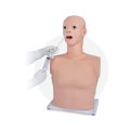
1. Life like, clear anatomical structure, include: head,neck, chest, real size lungs and stomach
2. Transparent abdominal wall, clear observation of internal organs
3. Oral/nasal cavity intubation
ㅤ① Inserting to trachea, air feeding, lungs will be inflated
ㅤ② Inserting to esophagus, air feeding, stomach will be inflated
4. Nasal feeding:When the gastric tube is inserted at 45-55cm, the simulated gastric juice can be drawn out
5. Gastric lavage:practice through oral/nasal cavity, stomach capacity up to 500ml