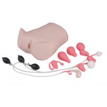
A comprehensive gynecological examination model is a lifelike replica used for training and practice in performing thorough examinations of the female reproductive system. This model includes anatomically correct structures such as the uterus, ovaries, cervix, and vaginal canal, providing a realistic representation of the female anatomy.
The purpose of this model is to enhance the skills and knowledge of medical students, residents, and healthcare professionals in conducting various gynecological examinations. Trainees can practice visual inspection, palpation, and the use of medical instruments in a controlled environment.
The comprehensive gynecological examination model is designed to simulate different conditions and abnormalities that may be encountered during actual examinations. It may include interchangeable parts or modules to replicate conditions like fibroids, cysts, or polyps. This allows trainees to learn how to identify and handle these conditions appropriately.

These models are commonly used in medical schools, training centers, and healthcare institutions for educational purposes. They offer a hands-on learning experience that helps trainees develop confidence, dexterity, and proficiency in gynecological examinations. Additionally, they can be used to educate patients about procedures, conditions, and treatment options by providing a visual and tactile demonstration.
In conclusion, a comprehensive gynecological examination model is an essential tool for medical education and training. It provides a realistic representation of the female reproductive system, allowing trainees to practice and improve their skills in conducting thorough gynecological examinations. The model can be used alongside other training models such as First Aid Skill Training Model, Full Body Trauma Manikin, and ACLS Training to provide a comprehensive medical education experience.
Features:
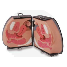
1. Female hypogastria, clear anatomical mark, lithotomy position
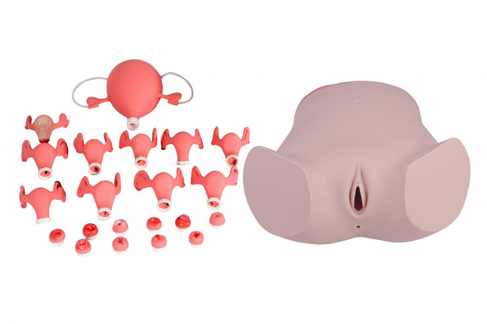
2. Palpation of normal and various abnormal uterines
3. Visual observation of normal and abnormal pathological changes of the cervix
4. Observe the size and position of the diaphragm
5. Bimanual examination
6. IUD placement and extraction
7. Observe anatomical structure of uterus, ovary, fallopian tube, etc.
8. Vaginal speculum and colposcopy examination
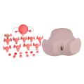
9. The components:
Normal and abnormal cervix (11pcs)
1. normal cervix
2. contraceptive device placing and extraction cervix
3. tearing cervicitis
4. chronic cervicitis
5. acute cervicitis
6. cervicitis bothe cysts
7. trichomonad sex cervicitis
8 . cervical condyloma acuminatum
9. leukoplakia of cervix
10. cervical polyp
11. Adenocarcinoma of the uterine cervix
Normal and abnormal uterus and accessories (11 pcs)
1. normal uterus and attachments
2. contraceptive device placing and extraction uterus
3. anteverted uterus
4. retroverted uterus
5. uterine fibroids
6. tubo-ovarian cyst (right side)
7. hydrosalpinx (right side)
8. tuberculosis of fallopian tube (right side)
9. uterus with salpingitis on right side
10. practice using contraceptive ring guide fork to place and extract contraceptive device (transparent)
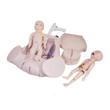
11. gravid uterus(fifth month fetus)