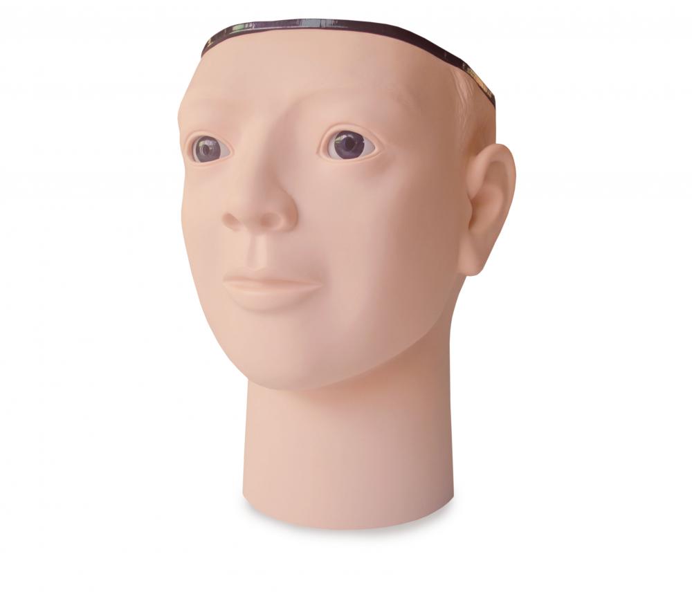
The retinopathy examination model is a valuable tool for training healthcare professionals in various medical fields. It allows for practice and refinement of examination techniques without the need for a live patient. Similar to other medical training models such as the First Aid Skill Training Model or the Trauma Nursing Model, the retinopathy examination model provides a hands-on learning experience.
 Using an ophthalmoscope, healthcare professionals can examine the model eye for abnormalities or signs of retinopathy. This process is similar to using an Intravenous Injection Arm or an Intramuscular Injection Model to practice injection techniques. The retinopathy examination model allows for a detailed examination of the retina, just as a Mattress Sutures Model enables practice in suturing skills.
In addition to training healthcare professionals, the retinopathy examination model can also be used for patient education. Just like an Aed Training or an ACLS Training model allows for simulated emergency situations, the retinopathy examination model helps healthcare professionals explain and demonstrate the condition and its implications to patients.
Overall, the retinopathy examination model is a specialized tool in the field of ophthalmology, similar to other medical training models like the Full Body Trauma Manikin or the Electronic CPR Manikin. It assists healthcare professionals in accurately assessing and diagnosing retinopathy, leading to appropriate treatment and management of the condition.
Features:
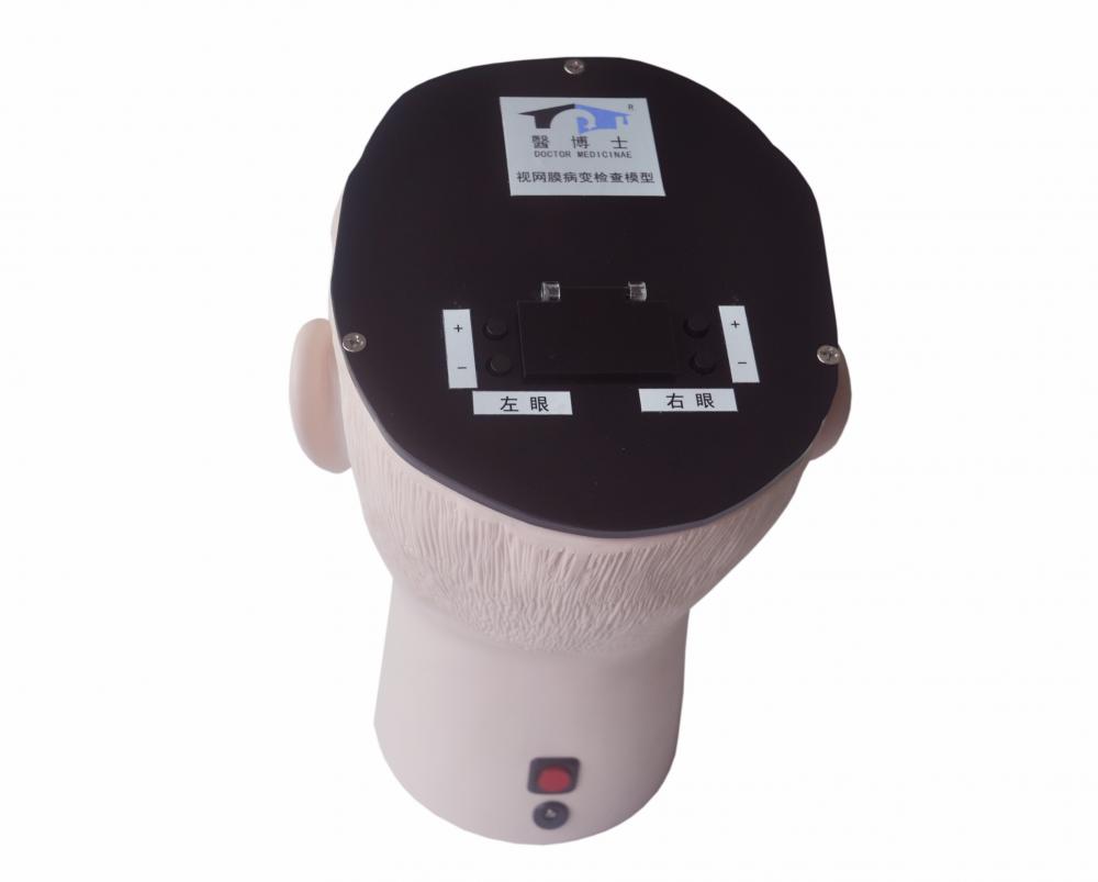
1. A well-shape adult head, electronic control
2. It simulates 29 retinopathies, left eye includes 17 types, right eye includes 12 types, switch to different retinopathies by press the button in back head
3. Left eye retinopathy, including:
1) Normal retina-2
2) Senile macular degeneration -2
3) Senile macular degeneration - glass warts-1
4) Central retinal vein occlusion
5) Hypertensive retinopathy-1
6) Optic nerve depression
7) Optic nerve atrophy-2
8) Background type diabetic retinopathy-1
9) Background type diabetic retinopathy-4
10) Background type diabetic retinopathy-5
11) Background type diabetic retinopathy-6—with Cotton-wool spot and blood spots
12) Proliferative diabetic retinopathy -2
13) Proliferative diabetic retinopathy -4
14) Proliferative diabetic retinopathy-5
15) Proliferative diabetic retinopathy-2—with New blood vessels and fibrous proliferation
16) Proliferative diabetic retinopathy-3—with New blood vessels, fibrous proliferation and concurrency retinal detachment
17) Systemic lupus erythematosus retinopathy
4. Right eye retinopathy, including:
1) Normal retina-1
2) Branch retinal vein occlusion
3) Hypertensive retinopathy-2
4) Optic disc edema-1
5) Optic disc edema-2
6) Optic nerve atrophy-1
7) Background type diabetic retinopathy-2—with red spot, retinal microaneurysm
8) Background type diabetic retinopathy-3—with microangioma and small hemorrhagic spot
9) Proliferative diabetic retinopathy -1
10) Proliferative diabetic retinopathy -3
11) Proliferative diabetic retinopathy -6
12) Proliferative diabetic retinopathy-1—with new blood vessels or vitreous hemorrhage