0 Piece,Product Price: US $ 0
| Quantity(Piece/Pieces) | 1 ~ 5000 | >5000 |
| Est. Time(days) | 7 | To be negotiated |
If you finish the payment today, your order will ship out within the delivery date.
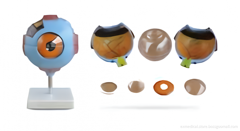
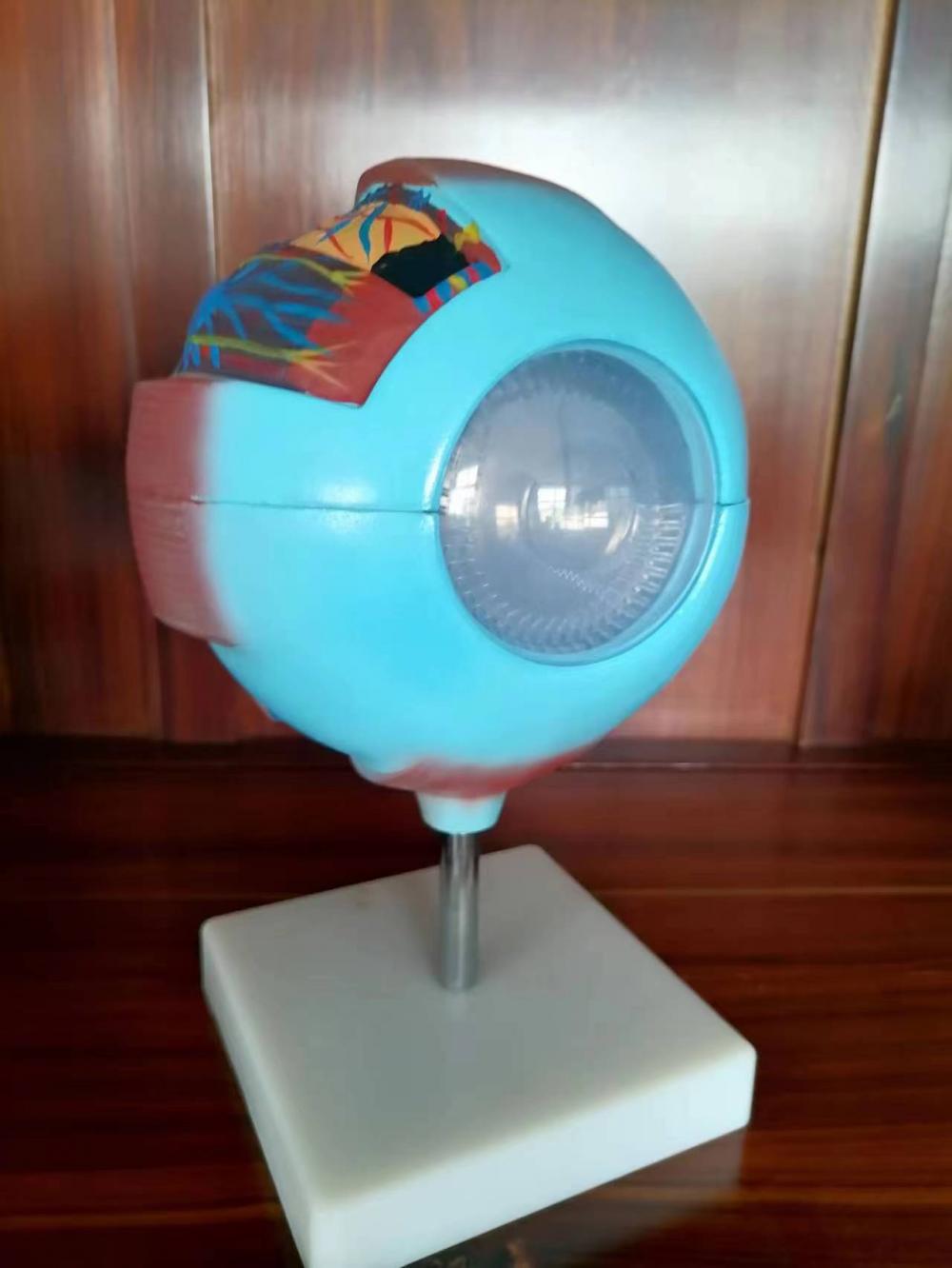
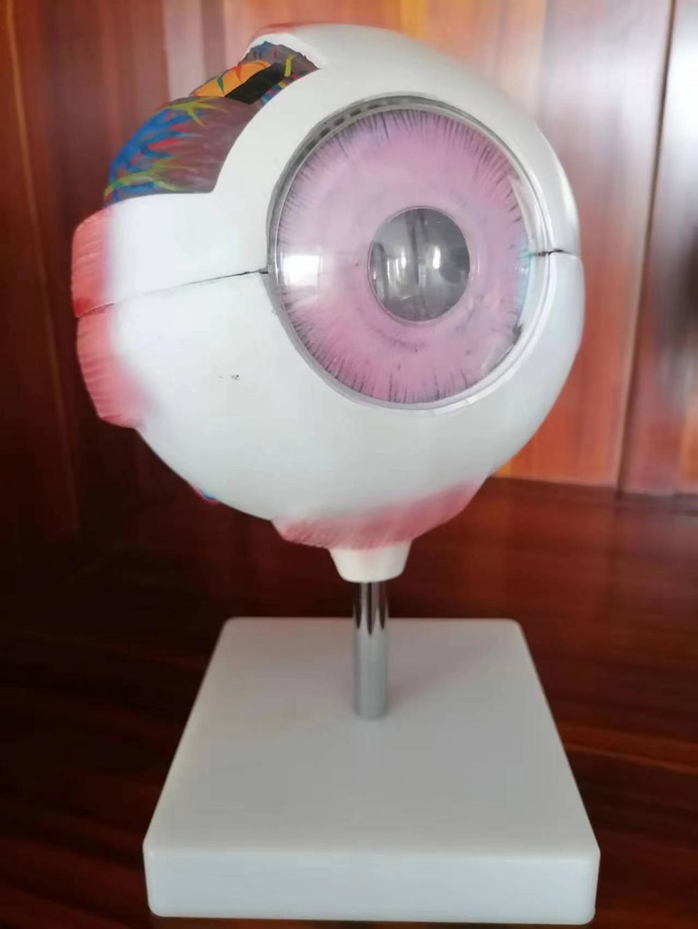
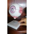
The human eye is a complex organ that enables us to see the world around us. It consists of several layers, including the outer layer, middle layer, and inner layer.
The outer layer is the most external part of the eye and includes the cornea and sclera. The cornea is a transparent dome-shaped structure that covers the front of the eye, helping to focus light onto the retina. The sclera is the white, tough outer layer of the eye that provides protection and support.
The middle layer is the intermediate layer of the eye and includes the iris, ciliary body, and choroid. The iris is the colored part of the eye that controls the amount of light entering the eye. The ciliary body is responsible for producing aqueous humor, which helps maintain the shape of the eyeball. The choroid is a network of blood vessels that supplies oxygen and nutrients to the retina.
The inner layer is the innermost layer of the eye and includes the retina, optic nerve, and vitreous humor. The retina is a thin tissue that covers the back of the eye and contains photoreceptor cells that convert light into electrical signals. The optic nerve is a bundle of nerve fibers that transmits these signals from the retina to the brain. The vitreous humor is a gel-like substance that fills the space between the lens and the retina, helping to maintain the shape of the eyeball.
To gain a more comprehensive understanding of the anatomy and function of the human eye, there are various training models and anatomical models available. These models are valuable tools for teaching and learning various medical procedures and skills. For example, there are models specifically designed for training in emergency medical skills, such as intravenous arm models and suture pad models. Additionally, there are full-body trauma models and high-fidelity simulation models for trauma training.
In the nursing field, there are models like trauma care models and advanced obstetric examination models that provide practice for specific nursing skills. Furthermore, there are models used for practicing diagnostic skills, orthopedic skills, surgical skills, obstetrics and gynecology skills, pediatric skills, endoscopy skills, nursing skills training, and specialized nursing.
For those interested in human anatomy, there are various human body models available, including skeletal models, nervous system models, sensory organ models, dental models, tooth models, and medical imaging models. These models provide visual representations of different anatomical structures, aiding in the study and understanding of how the human body works.
Overall, training models and anatomical models play a significant role in medical education, allowing healthcare professionals to develop and refine their skills in a safe and controlled environment. Whether it's simulating surgical procedures or examining anatomical structures, these models are crucial in medical training and education.
Features:
6 times enlarged eyeball demonstrates human eye anatomical structure, detachable for demonstrating tunica external, tunica media,
tunica internal and refraction media.


Worry-free return
Product quality protection
Payment protection
Shipping protection
Copyright © 2024 BOSSGOOMALL. All rights reserved
版权所有 宁波全贸信息技术有限公司 浙ICP备12012821号-84